Reading material on improving movement analysis and therapy
Every healthy individual performs approximately 20,000 respiratory movements within a day, primarily regulated by the diaphragm. This muscle is twice as vascularized as other striated skeletal muscles in humans and continuously functions from birth to death. Besides its respiratory role, the diaphragm plays a part in maintaining body posture, proper functioning of the vascular and lymphatic systems, and is involved in gastrointestinal activities such as swallowing, vomiting, acting as an anti-reflux barrier. The diaphragm influences the functioning of all abdominal organs. The interdependence of the diaphragm with various structures in the human body results in dysfunctions leading to discomfort in different areas of the body.
ANATOMY OF THE DIAPHRAGM
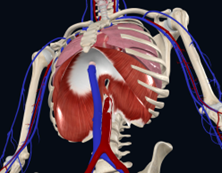
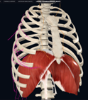
The diaphragm constitutes a transverse partition dividing the torso into the chest and abdominal cavities. It has the shape of a dome, with its upper part forming the floor of the pleural cavity, and its lower part creating the vault of the abdominal cavity.
The central part of this dome (the center of the diaphragm), situated highest in the chest, with fibers running horizontally, forms the central tendon composed of a fibrous layer that creates three leaflets (divisions): anterior (rich in lymphatic vessels), right lateral, and left lateral
The peripheral part of the dome consists of muscle fibers that are arranged radially around the chest. It is divided into sections:
- Sternal part: Connects to the xiphoid process of the sternum and the aponeurosis of the transversus abdominis muscle.
- Costal (lateral) part: Attaches to the cartilage of the sixth to seventh rib, interlocking with the attachments of the transversus abdominis muscle.
- Lumbar (posterior) part: Comprising two crura and arcuate ligaments:
- The right crus attaches to the L1-L4 vertebral bodies, intervertebral discs, and the anterior longitudinal ligament. During growth, the right crus gives branches to the esophageal hiatus and serves as a natural element functioning as the sphincter of the cardiac orifice of the stomach.
- The left crus attaches to the L1-L2 vertebral bodies and the anterior longitudinal ligament.
- The median arcuate ligament (lumbocostal arch) runs from the transverse process of L1 to the vertebral body, above the upper part of the psoas major muscle, connecting with its fascia.
- The lateral arcuate ligament (lumbocostal arch) extends between the transverse process of L1 and the apex of the 11th and 12th ribs. It covers the transverse process, then, connecting with the transversalis fascia, runs toward the abdomen, where it transitions into the pelvic fascia (Lierse 1990). The costolumbar hiatus is formed from this ligament.
Additional materials are available in the e-manual.
DIAPHRAGMATIC APERTURES
In the diaphragm, there are apertures through which structures from the abdominal cavity pass into the chest (Bochenek 1990).
Three main physiological openings:
- Aortic hiatus (descending aorta, thoracic duct): Located in the lumbar part, medially between the crura of the diaphragm and reinforced by the middle arcuate ligaments.
- Esophageal hiatus (esophagus, vagus nerves, branches of the left phrenic nerve): Located in the lumbar part in the right crus, entirely surrounded by muscles.
- caval opening (inferior vena cava and branches of the right phrenic nerve): Positioned on the right side, ventrally in the area of the central tendon.
Other anatomical openings:
- sternocostal triangle, Larrey’s space (superior epigastric artery and vein): Located in the sternal and costal parts.
- Lumbocostal triangle:
- Medial part (greater and lesser splanchnic nerves, azygos vein, and hemiazygos vein): Located in the lumbar part.
- Lateral part (sympathetic trunk): Positioned in the lumbar part, between the medial and lateral parts.
DIAPHRAGMATIC INNERVATION
- Phrenic Nerve: It is a mixed motor-sensory nerve originating from the anterior branches of spinal nerves C3-C5, which are part of the cervical plexus. Its primary function is to provide exclusive motor control of the diaphragm. It is a fundamental nerve in the physiology of respiration, supplying the sensory (pain and proprioception) part of the central diaphragm and adjacent fascial layers. The phrenic nerve is bilateral, but left and right nerve have some significant differences in terms of course and relationships with surrounding structures.
- Intercostal Nerves: The peripheral muscular parts are innervated by intercostal nerves from T6 to T11. Intercostal nerves are mixed nerves, carrying both motor and sensory fibers. Their main function is the segmental supply of structures in the chest wall and abdominal cavity. In addition to motor innervation of intercostal muscles and the anterolateral abdominal wall muscles, they carry sensory fibers conveying information from the skin, chest and abdominal wall, ribs, pleura, and peritoneum. These nerves also transmit sympathetic innervation to structures (sweat glands, blood vessels) in the chest and abdominal wall.
Diaphragmatic innervation is multi-neural:
- Central tendon (originating from the transverse septum): Phrenic nerve.
- Muscular momes (partially originating from the transverse septum and partially from the muscular walls of the trunk): Intercostal nerves innervate the lateral portion.
- The crura of the diaphragm (originating from the dorsal attachment of the esophagus – vagus nerve).
Additional materials are available in the e-manual.
DIAPHRAGM POSITION. REFERENCE POINTS
The position of the diaphragm depends on various factors, including body type, respiratory phase (inhale or exhale), depth of breath, and body position (standing, sitting, lying down).
On the upper side of the diaphragm, the central tendon is fused with the fibrous pericardium. The diaphragmatic domes rest over the lower lobe of the right lung and on the over the left lung (right and left dome respectively). On the lower side, the diaphragm is fused with the liver on the right, forming the bare area of the liver, and on the left, it is connected to the stomach and spleen.
When examining a patient in a sitting or standing position, the right diaphragmatic dome is slightly higher than the left, with a difference of 1 to 2 cm. In a healthy individual, the right dome extends to the level of the upper border of the fifth rib, while the left dome reaches the lower border of the sixth rib. During maximum exhalation, the diaphragm rises to the level of the fourth intercostal space, and during full inhalation, it flattens, lowering the chest cavity to the level of the rib arches anteriorly and the twelfth rib posteriorly.
The aortic hiatus is located to the left of the midline at the level of the T12-L1 vertebrae, and just inferiorly at the T10 level, the esophageal hiatus is found. At the level of T9 to the right of the midline, there is the opening of the inferior vena cava and the sternocostal triangle.
The median lumbocostal slit is located at the level of L1, and the lateral slit is at the level of L2.
It’s important to note that some of the above reference points may undergo slight variations depending on the patient’s body structure, activities performed, and positions adopted.
FUNCTIONS OF THE DIAPHRAGM
The diaphragm serves various crucial roles in the body:
- Respiratory function: the diaphragm plays a vital role in breathing, satisfying approximately 80% of the body’s oxygen demand.
- Enhancement of spinal stability: contraction of the diaphragm contributes to increased intra-abdominal pressure, thereby improving the stability of the spine.
- Improvement of motor control of the spine.
- Facilitation of fluid flow.
- Barrier to microbial spread: it acts as a barrier, preventing the spread of microorganisms between two body cavities.
- Activation of abdominal organs: through the fascial system, the diaphragm activates abdominal organs by transmitting the force of its contraction to individual organs.
All the functions of the diaphragm in the human body are not fully understood, and ongoing medical research continues to explore and deepen our understanding of its multifaceted roles.
CLINICAL SYMPTOMS OF DIAPHRAGM DYSFUNCTION
Clinically, symptoms of diaphragm dysfunction may manifest as:
- Pain or tension in the thoracolumbar junction
- Pain below the costal arch
- Postural Disturbances
- Respiratory system disorders: conditions affecting the respiratory system, including bronchitis, bronchial asthma, and sinusitis.
- Gastrointestinal system disorders: conditions related to the gastrointestinal system, as the abdominal organs have direct or indirect ligamentous connections with the diaphragm.
- Peripheral circulation disorders of the lower limbs: associated with pathology of the inferior vena cava and abdominal aorta.
- Lymphatic circulation disorders: including swelling in the lower limbs and abdomen.
- Urinary-genital system disorders: the kidneys have a direct relationship with the diaphragm.
- Instability in the L5-S1 vertebrae.
- Developing disc herniation.
- Diaphragmatic hernia: symptoms may include heartburn, eructation, and substernal pain.
- Signs of quadratus lumborum muscle weakness.
- Manifestations of iliopsoas muscle weakness.
Respiratory insufficiency can cause:
- Autonomic imbalance: compression of the vagus nerve leading to disturbances in organ trophics.
- Variable activity in the craniosacral rhythm: each phase of respiration influencing the activity of specific cranial bones.
- Esophageal widening: resulting in pressure on the cardiac portion of the stomach, disrupting its main function of breaking down complex proteins into simple amino acids, leading to impaired protein digestion.
- Decreased tone of the lumbar-pelvic muscle (incorporated in the diaphragm crura): leading to nephroptosis.
- Instability in the cervical spine: overloading the central part of the cervical spine, compression of the phrenic nerves.
Additional materials are available in the e-manual.
Breathing is a continuous activity accompanying humans throughout the day. If incorrect movement patterns emerge in the body, coupled with the continuity of this process, compensations in the musculoskeletal system develop, leading to disruptions in postural patterns.
The process of diagnosing and treating diaphragm dysfunction should commence with an assessment of the patient’s breathing pattern in a lying and/or sitting position. In the presence of observed dysfunction, the following steps are recommended:
– determine at which level and which structures, besides the diaphragm, are participating in the dysfunction.
– choose a suitable relaxation technique for the diaphragm and/or the organ involved in the diaphragm dysfunction.
– treat tense muscles in the neck, chest, or abdomen that accompany diaphragm dysfunction.
Assessment of respiratory movement is conducted in areas where various types of tissues and organs overlap. The following regions are evaluated:
- Transition between the chest and neck area
- Transition between the abdominal cavity and chest
- Area above and below the diaphragm:
The objective of the examination is to assess the behaviour of specific structures, encompassed by the therapist’s hands, during normal physiological breathing. Initially, direct palpation is applied to the observable elements, followed by the assessment of gliding movements of deeper tissues and organs.
After conducting an assessment and identifying areas with reduced or absent tissue glide, targeted therapeutic techniques should be applied to those specific areas to improve their mobility.

Leave a Reply
Want to join the discussion?Feel free to contribute!