Reading material on improving movement analysis and therapy
WHAT IS CORE?
Core stability encompasses the lumbo-pelvic complex and refers to the ability to maintain the balance of the spine within its physiological limits while preserving structural integrity. Several authors have proposed a more functional perspective to describe the core as the foundation of the kinetic chain responsible for facilitating the transfer of torque and momentum between the lower and upper extremities for gross motor tasks of daily living, exercise, and sport. Core stability necessitates instantaneous changes by the central nervous system to elicit appropriate combinations and intensities of muscle recruitment for stiffness (i.e., stability) as well as mobility demands of the system. Core stability requires the activity of the central nervous system (CNS) to trigger the right combination and intensity of muscle recruitment for stability and mobility.
FUNCTIONAL CORE ANATOMY
Core stability involves a muscular cylinder consisting of the transversus abdominis (TVA) and internal abdominal muscles at the front, the multifidus muscle and the rear fibers of the psoas major muscle at the back, the diaphragm on top, and pelvic floor muscles at the bottom. These muscles are anchored to the thoraco-lumbar fascia and the spine area, creating what’s known as the kinematic chain. One of the parameters influencing spinal mechanics and stiffness is intra-abdominal pressure (IAP).
Intra-abdominal pressure is increased through the tension of the thoracolumbar fascia and the contraction of muscles like the transversus abdominis and multifidus.
The heightened tension of the thoracolumbar fascia and the resulting intra-abdominal pressure, which stem from muscle contraction, also play a role in supporting each spinal vertebra within the region.
The lumbopelvic-hip complex, often referred to as the “core,” consists of the lumbar vertebrae, the pelvis, the hip joints, and both active and passive structures that either enable or restrict movement in these segments. The stability of any system refers to its capacity to limit displacement and preserve structural integrity. Hence, core stability can be defined as the ability of the lumbo-pelvic-hip complex to prevent the vertebral column from buckling and restore it to equilibrium following perturbations.
The core stability kinematic chain, which includes the iliac crest, the trunk, and the pelvic girdle, is responsible for postural control, overall movement control, as well as the distribution and transfer of forces in the lower limb area. The Panjabi model elucidates the mechanisms of central stabilization, consisting of three interconnected subsystems: passive (bony and joint structures), active (muscles), and the neural component (muscle control). Maintaining stability requires continuous interaction among all three subsystems.
Hodges and Richardson have described a “feedforward” mechanism, which means that deep muscles are activated before movements of the upper or lower limbs. This early activation ensures the stability of the trunk during these movements. The “feedforward” process involves recruiting deep muscles to support overall movement, allowing for smooth, uncompensated movements. In healthy individuals, the transversus abdominis and multifidi contract before the shoulders or legs move, stabilizing the lumbar spine.
Functional motion is defined as the ability to maintain balance, mobility, and stability along the kinematic chain. Integral functional movement is a model of precision and efficiency. Deficits in postural control, impaired balance, altered proprioception, and inefficient motor control contribute to pain, dysfunction, and incorrect movement patterns.
Research results indicate that weak central stabilization can increase the risk of limb injuries in physically active individuals. Many authors describe various classifications of core stability in dynamic stabilization. These include local stabilizers (single-joint deep muscles) and global mobilizers (multi-joint superficial muscles). Gibbons and Camerford proposed a functional model that further divided global muscles into stabilizers and mobilizers. On the other hand, Behm et al. categorized global muscles into mobilizers and load-bearing muscles.
Additional materials are available in the e-manual.
CORE STABILITY IN THE PREVENTION OF TRAUMATIC INJURIES.
Core stability encompasses the passive structures of the thoracolumbar spine and pelvis, as well as the active involvement of the torso muscles. Stability relies on neuromuscular control of the torso in response to both internal and external forces, including those generated by distant body parts and expected or unexpected disruptions. According to the general definition in sports medicine literature, stability forms the foundation for dynamic trunk control, enabling the generation, transfer, and control of force and movement to further segments of the kinetic chain. Core stability is a fundamental element in regular athletic activities, involving integrated activation of multiple segments to provide force generation, proximal stability for distal mobility, and the creation of interactive moments.
As mentioned by the authors, even slight disruptions in proprioception and core neuromuscular control can influence the risk of lower limb injuries in active populations and the occurrence of Low Back Pain (LBP).
However, it’s worth noting that the concept of core stability has both proponents and critics, and scientists have expressed varying viewpoints on the topic in the existing literature.
Additional materials are available in the e-manual.
FUNCTIONAL DIAGNOSIS OF THE CORE
Functional diagnostics is an area of physiotherapy that deals with assessing the musculoskeletal system, including the evaluation of recruitment, strength, and endurance of the core muscles. It allows for identifying dysfunctions, neuromuscular control disorders, and, based on these findings, planning and implementing a physiotherapy program to improve overall function. Implementing proper diagnostics is key to creating an optimal therapeutic plan.
The simplest way to assess core muscle function is to evaluate the voluntary contraction of the transversus abdominis muscle. This involves palpating the transversus abdominis muscle in a medial direction and just below the anterior superior iliac spines, right next to the rectus abdominis muscle. This assessment is performed while the individual “draws in” without taking a deep breath. This initial examination helps identify improper recruitment and/or muscle function. In addition to palpation, objective methods like ultrasound (USG) are also used to assess muscle contraction. To evaluate core muscle strength and endurance, diagnostic tests such as the McGill Muscular Endurance Test protocol can be employed.
The test consists of 3 tests, and the results are recorded in a prepared protocol sheet.
- The Trunk Flexor Endurance Test evaluates the endurance of the deep abdominal muscles, including the transversus abdominis, quadratus lumborum, and erector spinae. It is also a timed, isometric test that focuses on the static contraction of these muscles, which help stabilize the spine, and continues until fatigue or significant compensatory movements of the torso occur. (photo 1)
- The Trunk Lateral Endurance Test measures the endurance of the lateral muscles, including the transversus abdominis, obliques, quadratus lumborum, and erector spinae. This timed test involves static, isometric contractions of the lateral muscles on both sides of the torso to stabilize the spine. The test is performed with the participant lying on their side, raising their hips, and supporting their body weight on the elbow and feet. This test is conducted for both sides of the body. (photo 2)
- The Trunk Extensor Endurance Test assesses the endurance of the back muscles, including the erector spinae, latissimus dorsi, iliocostalis, and multifidus muscles. It is a timed, isometric test that involves a static contraction of these extensor muscles, which stabilize the spine. The test is conducted with the participant lying face down, hips and upper torso extending over the edge of a table, while the lower limbs are stabilized. (photo 3)
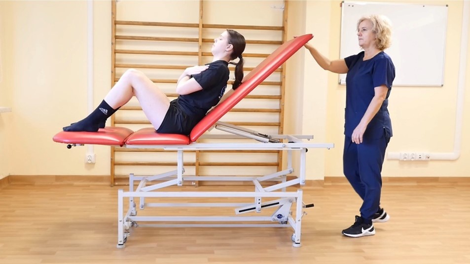
photo 1
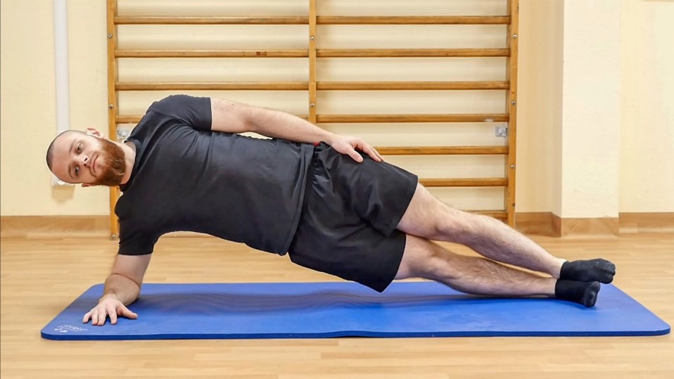
photo 2
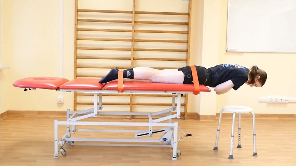
photo 3
The results obtained from the tests, which are measured in seconds, are entered into a prepared study protocol sheet and then subjected to analysis.
McGill’s torso muscular endurance test battery—record sheet
| Trunk flexor endurance test Time to completion: _______________ |
| Trunk lateral endurance test Right side time to completion: ___________ Left side time to completion:___________ |
| Trunk extensor endurance test Time to completion: _______________ |
| Ratio of Comparison Criteria for Good Relationship Between Muscles Flexion:extension Ratio less than 1.0 Right-side bridge:left-side bridge Scores should be no greater than 0.05 from a balanced score of 1.0 Side bridge (each side):extension Ratio less than 0.75 |
| Flexion:extension ratio: ________________ Rating: q Good q Poor |
| Right-side bridge:left-side bridge ratio: _________ Rating: q Good q Poor |
| Side-bridge (each side):extension ratio: _________ Rating: q Good q Poor |
https://docs.google.com/document/d/1iiVclYqIraPJsKZfOpvW_As0uD4vftkJg0Dm-qvgGnA/edit?usp=sharing
TRAINING
After conducting the functional diagnostic tests, such as the McGill protocol, and analyzing the results, a training or physiotherapy program can be planned. The intervention should be individually tailored to the exerciser’s fitness level. To increase the intensity of the exercises, progressive training should be applied. This includes upper body movements, diaphragmatic breathing exercises, unstable surfaces, and sport-specific functional training. Progressive exercises improve muscle recruitment and have a positive impact on sports performance and injury prevention.
Training the core muscles is an integral part of physiotherapy and forms the basis for healthy body movement. Dysfunctions in core stability can lead to increased forces on the spine and the development of compensations in the distal parts of the body. This, in turn, can result in spinal overloads and biomechanically inefficient movements, leading to reduced overall function.
Core stability training involves exercises for the muscular corset surrounding the torso and the lumbo-pelvic complex. Its primary purpose is to provide stability and protection for the spine during various movements, ranging from everyday activities to complex motions in sports.
This is linked to their function, which includes:
- Reducing forces acting on the spine.
- Facilitating the proper and most efficient transfer of forces from the lower part of the body to the upper part, and vice versa.
Endurance core stability is a vital element in preventing traumatic injuries and Low Back Pain (LBP), as well as enhancing athletic performance. Injury prevention is linked to better spine stability during movement and the ability to perform physiological movement patterns without pathological compensations. To achieve mobility in peripheral joints, it is necessary to establish proximal stability beforehand. An essential aspect of training is proper controlled diaphragmatic breathing, which determines the correct tension of deep muscles.
Core muscle training, like any other muscle training, should be progressive, meaning that the training load should be gradually increased. It’s important to remember that too rapid progress can lead to incorrect exercise patterns or compensatory movements.
Core exercises may include: Bird dog, Side plank, McGill sit-ups, Glute Bridge Hold, and Pallof press with a resistance band.

Leave a Reply
Want to join the discussion?Feel free to contribute!